


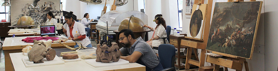
Documentation
Conservation documentation can be defined as the textual and visual records collected during the treatment of an object. It includes records of the object’s condition (condition assessment), the treatment done to the object and details on the object’s past and present environment. Good documentation should provide as much information as possible on the object for the future researcher, curator, or conservator.
There are many forms of documentation and these are not universally agreed upon. Digital technology has now made conservation documentation more easily accessible, cost/time efficient, and has also increased consistency and accuracy of the recorded data, and reduced physical storage space requirements.
Written documentation – Components of written documents which can be used as the markers for the identification includes. Documentation is further continued during the treatment procedure in conservation.
Graphical documentation is one of the methods to document the deterioration pattern of an object. It can be achieved either manually by drawing the object by free hand or by tracing or with the use of various software’s which can aid in copying the image of the object. Graphical documentation helps in knowing the object better as we have an image for reference and we can mark the areas where we see, inscription, stains, cracks, scratches and various other deterioration patterns.
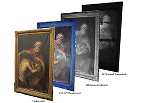
Photography is one of the important visual records of an object. While recording and documenting deterioration patterns it is important to take context image, detailed image and micro images for defining the area with deterioration to give a better understanding of the area to be treated. Photography combined with different angles of light such as raking light, transmitted light, and filters for light like infra-red, reflected or ultraviolet light, can give you more information than what the eye meets about the object
Multi spectral Imaging – A multispectral image is one that captures image data within specific wavelength ranges across the electromagnetic spectrum. The wavelengths may be separated by filters or detected via the use of instruments that are sensitive to particular wavelengths, including light from frequencies beyond the visible light range, i.e. infrared and ultra-violet. Multispectral imaging has also found use in document and painting analysis
Reflectance Transformation Imaging (RTI) is an Interactive versions of raking light photography that allow the point light source to be digitally manipulated by combining a sequence of images taken under varying conditions is an image-based representation of the appearance of a surface under varying lighting directions. Per-pixel surface normal are extracted from the representation, and can be used for not only changing lighting direction interactively, which can be read by RTI viewer.
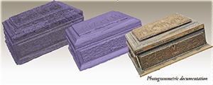
Photogrammetry is the art, science, and technology of obtaining reliable information about physical objects and the environment through processes of recording, measuring, and interpreting photographic images and patterns of recorded radiant electromagnetic energy and other phenomena. The fundamental principle used by photogrammetry is triangulation. By taking photographs from at least two different locations, so-called “lines of sight” can be developed from each camera to points on the object. These lines of sight (sometimes called rays owing to their optical nature) are mathematically intersected to produce the 3-dimensional coordinates of the points of interest.
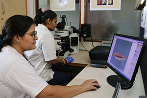
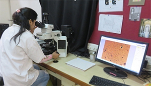
Microscopic studies
Thermal imaging is simply the process of converting infrared (IR) radiation (heat) into visible images that depict the spatial distribution of temperature differences in a scene viewed by a thermal camera. The imaging camera is fitted with an infrared detector, usually in a focal plane array, of micron-size detecting elements or “pixels.”
Microscopic studies
Examination is a detailed inspection or study of an object, to observe the general details of the object and to mark its distinctive traits, which is notified under description of the object, as well as to observe the deterioration pattern or extend of the damage occurred on the object. Examination can be classified based on the technique used as invasive and non-invasive.
Non – Invasive Methods of Examination
Visible light encompasses daylight, incandescent and fluorescent light sources. Examination with visible light is used in the initial stage of the examination of works of art. Such examination is frequently aided by the hand-held magnifiers or a stereomicroscope, to zoom in on features of special interest.
Raking Light When light source is located to one side of the object at low angle with respect to the examined surface, it is called raking or grazing light. This type of examination emphasizes the painted layer’s texture like brushstrokes, flaking paint and support deformations like dents, abrasions etc.
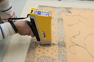
X-ray fluorescence is an analytical technique that returns information about the elemental composition of a sample. The sample is illuminated with an X-ray beam and the atoms which are struck by the beam emit X-rays in response, usually at several different energies.
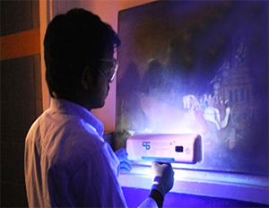
UV examination of artworks is a non-destructive technique that is extremely useful in the assessment of the surface of an object. Few inorganic materials and many organic materials do emit a visible fluorescence when irradiated with ultraviolet emissions. The study and documentation of this fluorescence can aid in the determination of the condition of an object, the extent of any over paintings or retouches and the presence of any resinous coatings.
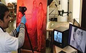
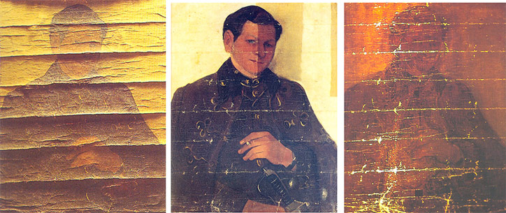
Infrared imaging is a non-destructive technique used by conservators to examine paintings and artworks and detect hidden details under the upper layers such as added paint, underdrawings, and hidden signatures or watermarks. It also can be used as a tool to differentiate between certain groups of pigments and inks
Invasive Techniques
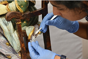
Sampling: Invasive examination mostly starts with taking a small sample from the body of the object for understanding the material constituents on the body of the object. The sample is examined unmounted under a microscope first before embedding it in a block. Samples are to be mounted the materials required, are carbon clay, a thin glass microscope slide and a high magnification microscope and then is embedded in a resin block and is further sanded to uncover the layers to study the cross-sectional area under a microscope.
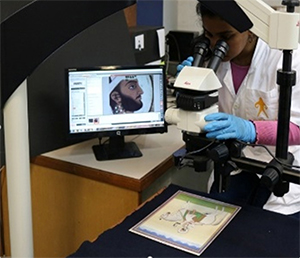
Microscopy is an important tool in effective conservation and restoration which aids in examination and research to understand the composition and condition of the artefact. From stereo microscope to electron microscopy, the optics used are dictated by the objects being examined.
Polarized light microscopy is another common technique used when identifying pigments is necessary. It involves analyzing very small paint chip samples of finish layers (right down to, and including, the substrate, so nothing is missed) and examining the cross sections for colors and material characterization.
Scanning Electron Microsopy(SEM) and Energy dispersive X-ray spectroscopy
(EDS, EDX)is used for imaging is good for elemental analysis of art works. SEM/EDS can be used for both organic and inorganic materials.
Fourier Transform Infrared Spectrometry is used to determine the type of paint (chemicals, pigments, etc.) by analyzing the way in which its various components absorb infrared light. The FTIR technique is useful for analyzing the chemical composition of smaller particles as well as larger areas on the surface. FTIR can be best used for the analysis of materials to aid in the choice of the optimum solvent needed to selectively remove certain coatings while leaving others unaffected.
The application of Raman spectroscopy in the field of non-destructive art analysis enables the identification of inorganic and organic pigments as well as binding media and varnishes